
Page 7 - 21st Century Perspective - Glaucoma Supplement
P. 7
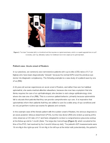
Figure 2. The Icare Tonometer (left) is a handheld unit that requires no topical anesthesia, which is a superb upgrade from air-puff
tonometry, and may ultimately replace the Goldmann tonometer (right) as the gold standard.
Patient case: Acute onset of floaters
In our practices, we commonly see normotensive patients with cup-to-disc (C/D) ratios of 0.7 (or
higher) who have been diagnostically “missed,” because the normal IOPs lured the previous eye
doctor into diagnostic complacency. The following example is a case study of a patient seen by one
of us (RM):
A 62-year-old woman experiences an acute onset of floaters, and rather than see her habitual
optometrist, she seeks medical attention elsewhere, because she now has a symptom that she
thinks requires the care of an ophthalmologist; she decides to visit a large ophthalmology clinic
where she saw one of us (RM). This is a common patient behavior, primarily because optometrists
fail to educate their patients that they can provide comprehensive eye care. It is imperative that
optometrists inform their patients that they are skilled to care for a wide array of eye conditions and
do not just perform routine eye exams for glasses and contacts.
In this example case of the female patient with the sudden onset of floaters, the obvious diagnosis is
an acute posterior vitreous detachment (PVD), but the new doctor (RM) she ended up seeing at the
clinic observes a C/D ratio of 0.7 and feels obligated to conduct a comprehensive glaucoma workup
at the follow-up visit in 1 month (Note: The large disc hemorrhage [Figure 3], which occurred at the
time of the PVD, had resolved by the 1-month follow-up visit). The patient’s IOP measurements were
18 mm Hg in the right eye and 19 mm Hg in the left eye at the initial visit (coincidentally, the patient’s
7